A pregnancy scan is not one interchangeable test: each examination is designed for a particular question and gestational window. By understanding what each scan checks, how to prepare, and how results change next steps, you can avoid missed screening windows and know when a delay needs prompt clinical advice.
Key takeaways
- Ultrasound uses sound waves, not ionising radiation.
- Book time-dependent scans within the recommended gestational window.
- A delayed scan can limit which measurements and screening assessments remain useful.
- Take your report to your obstetrician for a clear next-step plan.
What a pregnancy ultrasound can—and cannot—show
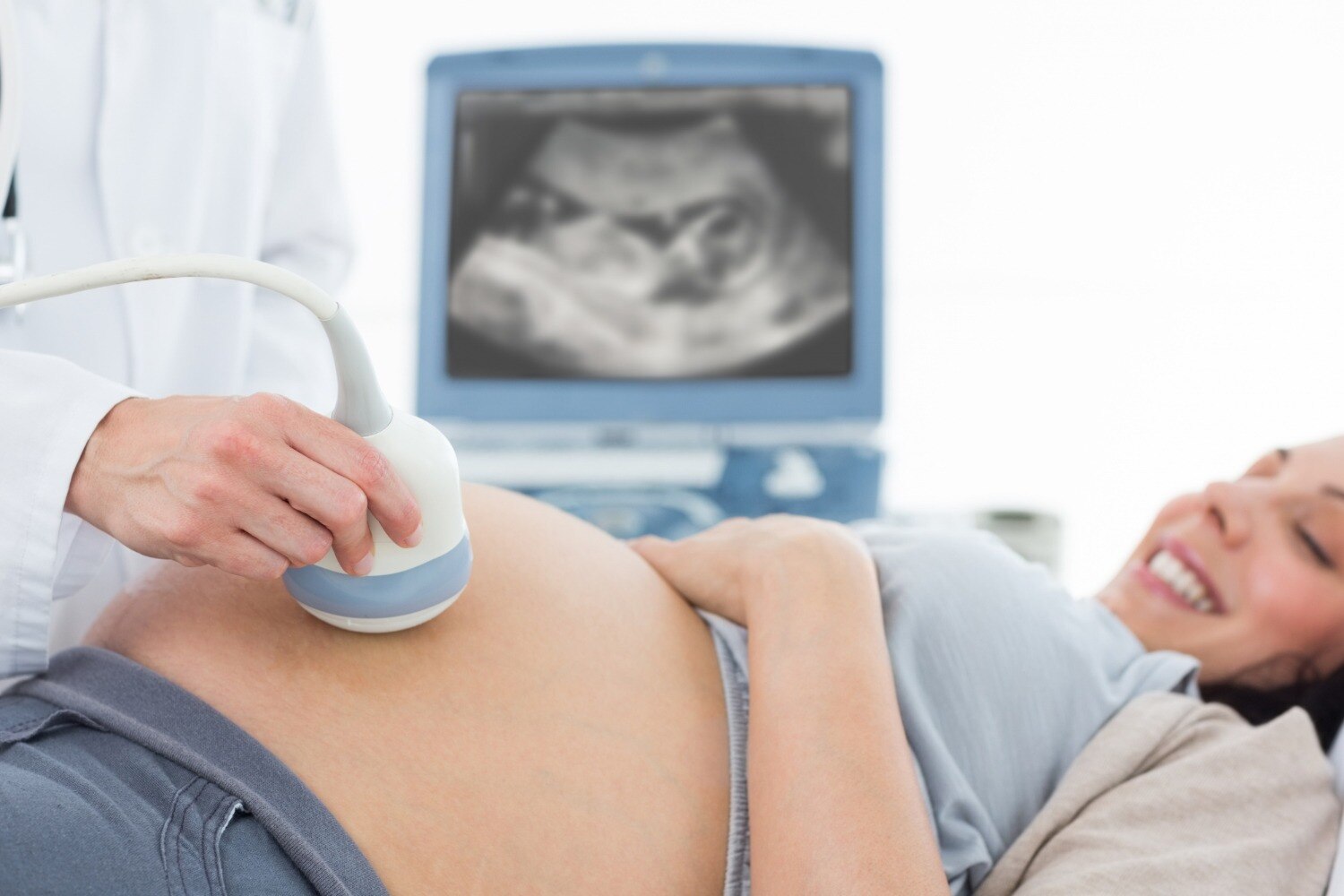
A pregnancy ultrasound examination uses a transducer placed on the abdomen or, in an early transvaginal scan, inside the vagina. The transducer sends and receives sound waves; software converts their echoes into moving and still ultrasound images produced in pregnancy. It uses no ionising radiation.
One early examination can assess several questions, but a very early scan cannot always answer all of them:
| Question | What the scan may show | Important limit |
|---|---|---|
| Pregnancy location | Whether the pregnancy is inside the uterus | A very early scan may not yet show a gestational sac |
| Cardiac activity | Whether embryonic or fetal cardiac activity is visible | No heartbeat at an early stage does not automatically mean miscarriage |
| Number of embryos | A singleton or multiple pregnancy | In twins, chorionicity and amnionicity are easiest to establish in the first trimester |
| Development | Whether measurements appear consistent with the estimated gestational age | Dates based on the last menstrual period can be inaccurate |
A routine scan surveys expected structures for that stage. A targeted diagnostic scan focuses on a specific concern, such as pain, bleeding, uncertain location or a suspected abnormality; a reassuring image cannot replace formal screening or diagnostic testing.
If the scan shows an empty sac or no heartbeat, clinicians use strict size and timing criteria and may arrange a repeat scan before diagnosing pregnancy loss.
Pain, vaginal bleeding, dizziness, fainting or shoulder pain needs prompt assessment rather than a routine appointment, because ectopic pregnancy can be life-threatening. A trained operator uses the lowest acoustic output and shortest exposure needed to answer the clinical question.
The pregnancy scans that depend on timing
The main pregnancy scans are timed to answer different questions, not to produce repeated reassurance. A scan done too early or late can leave a specific question unanswered.
- Dating and viability scan: Usually performed around 6 to 10 weeks, it checks whether the pregnancy is inside the uterus, whether cardiac activity is present, how many embryos are developing and whether size matches the expected gestational age.
Crown–rump-length dating is most accurate in the first trimester, especially when the last menstrual period is uncertain.
- Nuchal translucency scan: Performed from 11 weeks 0 days to 13 weeks 6 days, when the crown–rump length is about 45–84 mm, it measures fluid at the back of the fetal neck and assesses early anatomy as part of first-trimester screening. Missing this window changes which screening tests are available.
- Anomaly scan 18 to 22 weeks: This detailed examination checks the brain, spine, face, heart, abdomen, limbs and other organs. It also assesses placental position, amniotic fluid and fetal growth.
- Growth scan: Usually requested later in pregnancy when measurements, symptoms or risk factors raise concern. It asks whether the fetus is growing as expected and measures fluid volume.
- Doppler study: Used when blood-flow assessment is needed, it examines circulation in vessels such as the umbilical artery and can help assess placental function.
- Biophysical profile: Usually performed later when fetal wellbeing needs closer assessment. It combines ultrasound observations such as movement, breathing movements, muscle tone and amniotic fluid.
Why delaying a scan can change the information available
An ultrasound is a time-specific measurement, not a permanent record. Crown–rump length dating is most accurate in the first trimester, usually within about 5–7 days; later scans have a wider dating error. Changing an established due date because a late scan suggests a different age can therefore mislead decisions about screening, growth and delivery.
The nuchal-translucency window runs from 11 weeks 0 days to 13 weeks 6 days, with a crown–rump length of about 45–84 mm. Missing it does not end prenatal screening, but it changes which tests remain available.
In twins, early assessment of chorionicity and amnionicity is easier and more reliable; later uncertainty can make it harder to choose the right surveillance schedule.
A delayed scan can also change the next clinical decision without automatically requiring treatment. A growth scan showing a small estimated fetal weight needs interpretation alongside interval growth, fluid, maternal history, accurate dating and a Doppler study, because one measurement has meaningful error. The result may lead to:
- A repeat scan after an appropriate interval
- Closer blood-pressure or fetal surveillance
- Referral for specialist assessment or delivery planning
More scans are not automatically better in an uncomplicated pregnancy. If an early scan shows an empty sac or no heartbeat, strict size and timing criteria matter; a repeat scan can prevent a viable pregnancy from being wrongly diagnosed as a miscarriage.
What to expect before, during and after the examination
Preparation depends on whether the examination is abdominal or internal. Follow the instructions from the clinic, because the wrong bladder state can make early images harder to interpret.
1. For an abdominal ultrasound, drink water beforehand and arrive with a comfortably full bladder if the clinic requests one. The bladder creates a window through which the uterus and early pregnancy are easier to see. Do not overfill it to the point of pain.
2. For a transvaginal ultrasound, empty your bladder immediately before the examination. You undress from the waist down and lie on an examination couch. The clinician inserts a covered lubricated probe gently into the vagina; tell them if you feel pain so they can adjust the examination.
3. During either scan, the clinician moves the transducer to obtain views of the pregnancy, uterus, cervix, placenta and ovaries when needed. You may see images on the screen, but the person performing the scan may need time to record measurements before discussing them.
Afterward, you can usually return to normal activities. Ask when you will receive the written report and who will explain any follow-up. An empty sac or absent heartbeat in a very early scan is not automatically a miscarriage diagnosis; strict measurements and a repeat scan may be necessary.
Seek prompt medical assessment rather than waiting for a routine appointment if you have pain, vaginal bleeding, dizziness, fainting or shoulder pain.
Turning a scan report into the next clinical decision
A sonographer obtains the images, but a qualified radiologist, obstetrician trained in fetal imaging, or fetal-medicine specialist interprets them and signs the report. The report should state the gestational age used, key measurements, clinical findings, limitations and a clear impression—not simply attach pictures.
The pregnancy scan center should link each finding to a time-bound next step. A suspected growth problem needs interval repeat imaging, Doppler assessment, amniotic-fluid measurement and review of gestational-age accuracy, because one estimated fetal weight is not proof of fetal growth restriction.
A biophysical profile may support a decision about antenatal monitoring when reduced movements, abnormal Dopplers or another concern warrants closer surveillance.
- An empty gestational sac or absent heartbeat early in pregnancy: apply strict diagnostic size and timing criteria before diagnosing miscarriage; arrange repeat imaging when the criteria are not met.
- Pain, bleeding, dizziness, fainting or shoulder pain: direct the patient to prompt clinical assessment rather than a routine appointment; ectopic pregnancy requires examination and, when indicated, serial hCG testing.
- A twin pregnancy: record chorionicity and amnionicity early, then tailor surveillance because dichorionic and monochorionic pregnancies carry different risks.
For someone receiving care from Dr Suparna Ganguly, the center should send the signed report, images and any urgent recommendation directly to the treating team, while explaining what happens next to the patient. A reassuring keepsake image cannot replace formal screening, diagnostic testing or symptom assessment.
Frequently asked questions
What can a pregnancy ultrasound show?
It can produce moving and still images of the pregnancy, helping assess development and selected anatomical features. It cannot answer every pregnancy or health question.
Why does the timing of a pregnancy scan matter?
Certain scans and measurements depend on gestational age. Delaying them can move the pregnancy outside the window when specific information is most useful.
What happens before, during and after a pregnancy ultrasound?
You may receive preparation instructions, such as arriving with a full bladder for some early abdominal scans. During the examination, a transducer is placed on the abdomen or used transvaginally when indicated. Afterward, review the report with your obstetrician.
What should you do with a pregnancy ultrasound report?
Discuss the findings with your obstetrician, who can relate the measurements and observations to your pregnancy and decide whether routine follow-up, another scan or further assessment is needed.